Hacettepe University Faculty of Medicine
Basic Oncology Subdivision
Basic Oncology Subdivision is within the scope of Hacettepe University Cancer Institute which was established in 1982, with the purpose of fulfilling the need of basic and clinic levels on oncology and training the society on protective measures by performing epidemiological researches. Basic Oncology Subdivision was commissioned in 1982 under the presidency of Prof. Dr. Emin Kansu.
In 1993 it became a fully-equipped basic cancer researches center within the current physical structure of Hacettepe University Oncology Hospital. Basic Oncology Subdivision trains specialists and researchers who are able to carry out routine diagnosis services and researches on cancer biology and immunology. These services are offered by faculty members and instructors, research assistants and technicians.


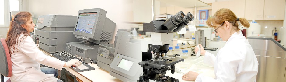
In Basic Oncology Subdivision, laboratory services on diagnosis and treatment of cancer and of certain diseases that effect immune system. In this context immune phenotyping (flow cytometry), cytogenetic and fluorescent in situ hybridization (FISH), tissue typing, immunology and cryopreservation methods are performed by a specialized team.
The diagnosis and classification of hematologic cancers are practiced by using flow cytometry, cytogenetic and FISH methods. In the meantime, the laboratory tests on mycosis fungoides, AIDS and other immunologic diseases that cause change in immune cell levels are also performed with flow cytometry method. In the tissue typing section, the PCR-SSP and PCR- SSO methods are used for genetic analyses of individuals who may be compatible donors for bone marrow transplantation patients in the section. For the peripheral blood stem cells (apheresis products) that are collected from these individuals and from patients, the procedure of gradually freezing at -196°C and preserving it in liquid nitrogen vapor phase (cryopreservation) is executed. Furthermore, cryofibrinogen and cryoglobulin designations are also among the services of Basic Oncology Subdivision.
Patients who will benefit the diagnosis services or samples taken from these patients are referred to the subdivision by the outpatient clinic and service physicians. Approximately 3000 patients benefit the diagnosis services of Basic Oncology Subdivision, in a year.
In hematologic cancers, flow cytometry method is benefited in evaluation of diseases by which the quantitative, physical and functional characteristics of immune cells. The presence of molecules on the surfaces of cells and their level changes are analyzed with this method. Diagnosis and determination of sub-types of leukemia and lymphoma (immunophenotyping), designation of lymphocyte subgroups and cell cycle-DNA analyses are performed.
How is the test performed?
Mononuclear cell suspension is prepared with peripheral blood and bone marrow samples. The panel to be proceeded is defined in accordance with the possible diagnosis of patient. For acute and chronic leukemia, the cells are marked with monoclonal antibodies that include lymphoid and myeloid cell markers. Then the cells are analyzed with flow cytometry in accordance with largeness, granularity and colors of marked monoclonal antibodies.
Considerations for flow cytometry test
For the test, 2-3 ml of blood or bone marrow samples are taken from patients into tubes with EDTA (purple covered) in the departments that they are examined. The maximum volumes of samples of other body fluids as far as possible (effusion, cerebrospinal fluid, etc.) should be taken.
Sample admission is proceeded on every weekdays, except Fridays, between 08:00 and 12:00. The patient should apply to Basic Oncology Subdivision, with the routing documents received from the departments that the patients are examined.
Test result can be received from the secretariat of relevant division via hospital system or from the Secretariat of Basic Oncology Subdivision, after 14:00 on the next day.
The designation cryoglobulin and criofibrinogen that are the proteins settle in cold is performed in immunology laboratory. These are the laboratory analyses that helps diagnosing vasculitis or glomerulonephritis diseases, in individuals who have circulatory problems on skin, particularly on arms and legs, when they are exposed to cold.
How is the test performed
Cryoglobulin designation is the method of defining monoclonal or polyclonal (immunoglobulin) proteins that settle when cooled in serum samples obtained from blood. Immediately after taken, the blood sample is kept at 37°C for 2 hours. Then it is centrifuged and the serum splits. It is kept at +4°C all night and the proteins settled in serum are evaluated.
Cryofibrinogen designation is the method of determining proteins that settle when cooled in plasma obtained from blood. Immediately after taken, the blood sample is kept at 37°C for 2 hours. Then it is centrifuged and the plasma splits. It is kept at +4°C all night and the proteins settled in serum are evaluated.
Considerations for cryoglobulin and cryofibrinogen analyses
For cryoglobulin test, 2 ml of blood sample is taken from patients into a simple, not including any anticoagulant (red covered) tube in the departments that they are examined. For cryofibrinogen test, 2 ml of blood sample is taken from patients into a tube with EDTA (purple covered), in the departments that they are examined. Immediately after the sample is taken, it should be carried in a thermos, which includes water heated until 37°C. Otherwise the sample is not admitted.
Sample admission is proceeded on every weekdays, except Fridays, between 08:00 and 12:00. The patient should apply to Basic Oncology Subdivision, with the routing documents received from the departments that the patients are examined.
Test result can be received from the secretariat of relevant division via hospital system or from the Secretariat of Basic Oncology Subdivision, after 14:00 on the next day.
In cryobiology laboratory, the procedure of long-term preserving the cells collected from patients with apheresis method by freezing for further use (cryoconservation) is performed.
The stem cells collected from patients who apply to Stem Cell Unit of Medical Oncology Subdivision and who are planned to have autologous or allogeneic bone marrow transplantation, are gradually frozen after adding chemicals that enables them to remain alive and kept in liquid nitrogen tanks at -196°C until the time of use. The procedure of thawing the frozen stem cells at 37°C when they will be administered to the patient, is among the services of Cryobiology Laboratory.
Tissue Typing Test
Tissue typing test, is performed to test the tissue compatibility between the patient (receiver) and the giver (donor) before bone marrow transplantation. The types of human leukocyte antigens, called HLA, are defined with this test. HLA antigens, exist on surfaces of almost all cells in human. Nervous (sunar ? sinir) and immune systems stimulate the foreign antigens that come into the body. HLA antigens have several types, however each person has certain number and types of HLA antigens. Some of them originated from mother ant the others are from father (genetic transition) and it is called "tissue type" or "HLA type". When the tissue transplantation is performed from a person whose HLA type is not similar, to another, the HLA type of donor may be perceived as "foreign" by the patient. In such a case, the immune system of the patient activates in order to destroy the transplanted tissue, thus the "tissue rejection" occurs. Tissue typing test is performed to find the most convenient donor that has the tissue compatibility with patient and to reduce the tissue rejection.
For whom is it applied:
Tissue typing test is performed in our laboratory before the bone marrow transplantations between relatives. It is applied firstly on the siblings, mother, father and children of the patient. If the compatible donor cannot be found, the 3rd and 4th degree relatives may also be scanned.
How is the test performed:
"Tissue Typing Test" is requested by the physician for the patient and his donors. The person who is requested to have test goes to the Blood Collection Unit and gives blood sample. The Sample Admission Section of Tissue Typing Laboratory of Oncology Hospital is applied along with the blood samples. The blood samples are received after the identification control is run. Patient Admittance Form is filled after a file is opened for the patient. The lines of descents and special conditions are recorded.
Tissue typing test is performed with molecular genetics methods. At first, the DNA isolation is practiced with the blood sample. The commercial kits and tests which include PCR-SSP and PCR-SSO methods are performed by using obtained DNA. The test results are analyzed and the report is prepared in compliance with HLA molecular genetics technology. The typing results of HLA-A, HLA-B, HLA-C, HLA-DQ, HLA-DR loci exist in the report.
Consideration for HLA typing analysis
Blood sample that is collected from the arm of the patient is used in tissue typing test. It is necessary to take the blood sample into two tubes with EDTA (purple covered).
The laboratory official should definitely be informed if blood transfusion takes place within last 15 days or if pregnancy is the subject matter. The sample is rejected if label information are not correct or deleted, if samples are clotted, if there are leakage, breakage or cracking on the sample tube, if the samples are not taken into the correct tube and if the information about patient or donor are incomplete (diagnosis of patient, name-surname of the physician who made request, address, etc.).
The results of tissue typing test are reported and submitted within 20 days. Reports are delivered to the physician of the patient inside a sealed envelope.
Since the reports include personal genetic information, they are not recorded into the information management system of hospital. They are protected in the computer of laboratory with passwords, no one, except the authorized laboratory officials can access them.
Conventional Cytogenetic and FISH Tests
By means of Conventional cytogenetic and Fluorescent In Situ Hybridization (FISH) methods, the structures called chromosomes in which the genetic information of each cell are kept are structurally and numerically analyzed (karyotyping). Hematologic cancers being in the first place, the chromosomal disorders that are determinants on diagnosis and follow ups of diseases are defined with karyotyping.
How is the test performed:
The bone marrow samples are reproduced for 24 hours in a sterile culture media that enables simultaneous reproduction of cells (synchronous culture). Then the collected cells are taken onto the lamina, analyzed and the chromosomes are enabled to senesce in an appropriate media. The chromosomes spreading by expanding are dyed (Giemsa-Trypsin banding method) and prepared for microscopic examination. The samples prepared in this way are examined structurally and numerically in the image analysis system and karyotyping is performed.
In FISH method, similar to the cytogenetic test, synchronous culture is generated with bone marrow samples. Following this procedure, the cells are collected and DNA particles (probes), marked with fluorescent that recognize the DNA regions to be analyzed, are applied. The probes are waited to connect to DNA and the unconnected particles are removed by washing. The cell nucleuses are also dyed (Dapi staining) and analyzed on image analysis system. Cytogenetic and FISH results are reported in compliance with "International System for Human Cytogenetic Nomenclature".
Consideration for cytogenetic and FISH analyses
Cytogenetic and FISH tests are performed through bone marrow samples. The samples that are taken into the heparin containing injectors or tubes (green covered) in sterile conditions should be delivered to the laboratory of Basic Oncology Subdivision within the shortest time, in order to minimize the cell loss. In addition, it is necessary for the bone marrow samples not to include clots, for the accuracy of test. Thus, after the sample is taken, it is recommended to turn the injector up and down for a few times, for the sample to be mixed with heparin. Otherwise the sample is not admitted.
Conventional cytogenetic result reports within 2-3 weeks and FISH reports within 10 days, can be received from the secretariat of division that the patient is examined by means of hospital system or from the Secretariat of Basic Oncology Subdivision.
Patients benefit the routine diagnosis services of Basic Oncology Subdivision through the requests and routings of the departments where they are examined. The blood samples should be delivered to the Secretariat of Subdivision (for tissue typing analyses to the Tissue Typing Laboratory - Secretariat of Apheresis Unit) along with the appropriate referral papers.
Secretariat of Subdivision, offices of faculty members and Basic Oncology Laboratories (Flow Cytometry Laboratory, Immunology Laboratory, Cytogenetic and FISH Laboratories) are located on the floor 1 of Hacettepe University Oncology Hospital. Tissue Typing Laboratory, Cryobiology Laboratory and Cytogenetic Analysis Section are located on the ground floor, within Apheresis Unit of Oncology Hospital.
Telephone:
+90 (312) 305 43 22, 0-312-3054323 (Secretariat of Subdivision)
+90 (312) 305 44 18, 0-312-3054325 (Routine Diagnosis Laboratory)
+90 (312) 305 44 11 (Flow Cytometry Laboratory)
+90 (312) 305 23 70, +90 (312) 305 23 72, +90 (312) 305 23 73 (Tissue Typing Laboratory)
+90 (312) 305 44 12 (Cytogenetic and FISH Analysis Section)